Menu ccc

PROTEIN EZ-VISION®, 4X

Cat.No. Size Price Quantity Subtotal Buy
N836-KIT KIT 來電價

Grade: BIOTECHNOLOGY GRADE

Products Description

Overview

  • Immediate visualization 
  • Sensitivity down to 100 ng protein
  • Compatible with downstream Western blotting   
 

Detailed Description

Protein EZ-Vision® is non-hazardous, flourescent reagent that produces instant visualization of protein bands upon UV illumination of SDS-PAGE gels. Supplied in a 4X loading buffer, Protein EZ-Vision® comigrates with the protein-SDS complex during electrophoresis. Post-run staining and destaining is completely eliminated and results can be visualized immediately after the run by placing the gel on a standard UV transilluminator. Available in 2 x 1 ml tubes (KIT) or in a 0.2 ml sample size.

Click on the Information tab to learn more about Protein EZ-Vision®, 4X

Information

Non-toxic alternative to Coomassie® staining


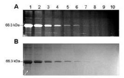
Sensitivity of Protein EZ-Vision® (A) compared to Coomassie® Blue sensitivity (B). Both figures were generated using the same gel comparison. Two-fold serial dilutions of bovine serum albumin (BSA) were separated via a 12.5% Laemmli SDS-PAGE gel using Protein EZ-Vision®, 4X as the sample loading buffer. Lane 7 is approximately 100ng protein. Immediate fluorescent visualization of protein post-electrophoresis was achieved with a 2 sec UV exposure using a SYBR® Green filter with the Syngene GBox-HR Gel Doc System (A). The same gel was post-stained with Coomassie® Blue protein stain for one hour and destained one hour (B).

Protein EZ-Vision® is compatible with downstream Western blotting applications.10μL of AMRESCO's Wide Range Protein Molecular Weight Markers (K494, lanes A1 and B3) and 10μg of K562 cell lysate (lanes A2 and B4) were run on a 12.5% Laemmli SDS-PAGE gel using Protein EZ-Vision®, 4X (N836) as the sample loading buffer. Immediate fluorescent visualization of protein post-electrophoresis in lanes A1 and A2 was achieved with a 2 sec UV exposure using a SYBR® Green filter with the Syngene GBox-HR Gel Doc System. Lanes B3 and B4 were post-stained with the Coomassie® Blue protein stain for one hour and destained one hour. Proteins from lanes A1 and A2, which were detected using Protein EZ-Vision® (A), were transferred onta a PVDF membrane with a semi-dry transfer unit and probed with Vimentin antibody using AMRESCO's Rapid Western Blotting Kit-Rabbit-SDT (N791, lanes C1 and C2). AMRESCO's VisiGlo Plus™ HRP Chemiluminescent Substrate Kit (N219) was used for detection.